Glymphatic System
Throughout most of the body, a complex system of lymphatic vessels is responsible for cleansing the tissues of potentially harmful metabolic waste products, accumulations of soluble proteins and excess interstitial fluid. But astonishingly, the body’s most sensitive tissue –the central nervous system – lacks a lymphatic vasculature. What then accounts for the efficient waste clearance that must occur in order for the neural tissue of our brains to function properly?
This question has puzzled scientists for centuries. Our group believes that understanding how this process functions in the healthy nervous system holds the key to developing treatment options for a wide variety of neurological diseases, especially those characterized by the improper accumulation of misfolded proteins. The breakdown of the brain’s innate clearance system may in fact underlie the pathogenesis of neurodegenerative disorders such as Alzheimer’s, Parkinson’s, and Huntington’s disease, in addition to ALS and chronic traumatic encephalopathy. Past efforts to explain how the brain cleanses parenchymal tissue have suggested that solute and fluid exchange occurs between the interstitial fluid and the cerebrospinal fluid, and that this exchange is driven by diffusion. Yet as many have noted, the distances for diffusion in the brain are too great to explain the highly regulated interstitial environment.
Recently, our lab has developed a fundamentally new approach to probing how the highly sensitive tissue of the CNS maintains its delicate extracellular environment. Our team made its initial discovery by injecting fluorescent tracers into the brains of living mice, and then imaging the movement of those tracers, in real time, using two-photon microscopy. These techniques allowed us to visualize the path of different sized molecules as they traversed the cortical layers of the brain, in addition to quantifying the clearance rate. Following the tracers revealed a distinctive pathway: once injected into the subarachnoid cerebrospinal fluid (CSF), the tracers readily flowed into the brain along the outside of the penetrating blood vessels. Further investigation confirmed that small channels 'piggybacking' the blood vasculature allow the CSF to flow into the brain tissue along para-arterial spaces and exit via a para-venous route. However, a critical component was still missing - based on diffusion alone, it would take over 100 hours for large solutes to traverse the inter- arterial and venous tissue.

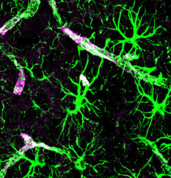
Astrocytes (green) with AQP4 expression on astrocytic end-feet (purple), surrounding cerebral vasculature (white).
The final puzzle piece came in the form of astrocytes, or more specifically, long projections they extend that envelop the brain vasculature. These 'end-feet' densely express the water channel aquaporin-4, which allow astrocytes to act as intervening 'couplers' of a bulk interstitial fluid (ISF) flow. Transgenic animals that lacked aquaporin-4 exhibited a 70 percent reduction in the clearance of large solutes, such as Amyloid-β peptide, whose extracellular plaques are the hallmark of Alzheimer’s dementia. This 'peri-vascular' route for CSF-ISF exchange constitutes a complete anatomical pathway, which we dubbed the glymphatic system
due to its dependence on glial cells performing a 'lymphatic' cleansing of the brain interstitial fluid.
Further characterization of the involvement of the glymphatic system in a number of neurological diseases and neurodegenerative disorders is underway. As an important further step to verify that the glymphatic system is functional in human brains, we are working in collaboration with researchers at the Stonybrook University to use contrast magnetic resonance imaging (MRI) to map regions of high and low volume solute exchange. Given that MRI is a commonly used clinical modality, it is our hope that this data may potentially enable the assessment of neurodegenerative disease risk and progression.
This project is supported by funding from NIH – National Institutes of Neurological Disorders and Stroke. (The Glymphatic System, a New Concept in Glia Biology
).
Further Reading
Evaluating glymphatic pathway function utilizing clinically relevant intrathecal infusion of CSF tracer. Yang L, Kress BT, Weber HJ, Thiyagarajan M, Wang B, Deane R, Benveniste H, Iliff JJ, Nedergaard M. J Transl Med. 2013 May 1;11(1):107.
Brain-wide pathway for waste clearance captured by contrast-enhanced MRI. Iliff JJ, Lee H, Yu M, Feng T, Logan J, Nedergaard M, Benveniste H. The Journal of Clinical Investigation. 2013;123:1299-1309.
A Paravascular Pathway Facilitates CSF Flow Through the Brain Parenchyma and the Clearance of Interstitial Solutes, Including Amyloid β. Iliff JJ, Wang M, Liao Y, Plogg BA, Peng W, Gundersen GA, Benveniste H, Vates GE, Deane R, Goldman SA, Nagelhus EA, Nedergaard M. Science Translational Medicine. 2012;147:1-11.


