If you're curious and passionate about science, then you might want to join us. We are always on the lookout for talented and motivated researchers to be part of our team. We have opportunities for all levels of education - enthusiasm and curiosity are the only requirements.
Depending on one's particular field of research, the next step is to send an email to the Principal Investigator with whom one would like to work, in order to present not only one's idea, but also oneself.
Background: Astrocytes are the primary homeostatic cells in the central nervous system (CNS), and one of their critical roles includes the regulation of extracellular ionic composition (ionostasis). Recently, we discovered that astrocytes serve as a dynamic reservoir for chloride (Cl-), significantly modulating inhibitory transmission that depends entirely on this anion. In a series of experiments involving awake, behaving mice, we observed that an increase in intracellular Cl- concentration in astrocytes ([Cl-]i) suppresses neuronal activity, while a decrease in astrocytic [Cl-]i enhances it. We provided evidence that this mechanism is mediated by Cl- efflux through GABAA receptors (GABAAR) expressed on astrocytes. Moreover, we found that astrocytic [Cl-]i is brain state-dependent, maintaining high and stable levels during sleep, and exhibiting dynamic, lower levels during wakefulness. These fluctuations in astrocytic chloride significantly influence the excitation/inhibition balance in both healthy and diseased brains. Next, we will investigate the implications of modulating astrocytic Cl- on sleep architecture and in pathophysiological conditions such as epilepsy.
The position: We are looking for a Master student to complete our team and help us decipher the role of astrocytic Cl- as regulator of E/I balance in sleep architecture or pathophysiological conditions such as epilepsy. We are using a broad spectrum of state-of-the-art imaging and electrophysiology techniques in in vivo mouse experiments.
About the lab: You will be part of a young team, which was founded in Nov 2022. Our lab consists of two Postdocs, and four PhD students. Our group is part of the Center for Translational Neuromedicine, which is part of SUND KU and located at Panum.
Your background: You are a highly motivated student with an interest in how the brain works. You are open and curious, eager to learn new techniques, a team player and interested in becoming a member of a young and dynamic, international team. Pre-requisites are good communications skills in English and a FELASA certificate.
Supervisors:
Verena Untiet, Associate Professor/Group Leader, Center for Translational Neuromedicine, KU
Zuzanna Bojarowska, PhD Student, Center for Translational Neuromedicine, KU
Katharina Baumgart, PhD Studnet, Center for Translational Neuromedicine, KU
Relevant Literature:
Untiet V, Verkhratsky A (2024) How astrocytic chloride modulates brain states. Bioessays DOI: 10.1002/bies.202400004
Untiet V, Nedergaard M, Verkhratsky A (2024) Astrocyte chloride, excitatory-inhibitory balance and epilepsy. Neural Regen Res DOI: 10.4103/1673-5374.390981
Untiet V (2024) Astrocytic chloride regulates brain function in health and disease. Cell Ca DOI: 10.1016/j.ceca.2024.102855
Untiet et al. (2023) Astrocytic chloride is brain state dependent and modulates inhibitory neurotransmission in mice. Nat Commun DOI: 0.1038/s41467-023-37433-9
Does this sound interesting to you? Just send an email to verena@sund.ku.dk, and we will schedule an informal meeting.
Have you ever wondered why it's challenging to get a good night's sleep on a warm summer night? Sleep is sensitive to temperature changes, and with climate change affecting our planet, maintaining quality sleep becomes a concern.
Quality sleep is crucial for cognitive performance and memory, yet the exact mechanisms remain mysterious. Sleep involves different stages, with Non-Rapid Eye Movement (NREM) being the most prevalent. NREM exerts its function through oscillatory patterns like spindles and Sharp Waves and Ripples (SWR) which are essential for memory and cognition.
On the other hand, neuromodulators like Norepinephrine and Acetylcholine play key roles in sleep stages, affecting arousal, memory, learning, and cognition. Recent research links EEG characteristics to these neuromodulators, showing their influence on the timing of spindles and SWR during NREM.
Our project aims to explore how changes in ambient temperature impact neuromodulator dynamics and neuronal oscillations. Using advanced methods like fiber photometry and traditional techniques such as local field potential recordings in mouse models, we'll investigate these effects in both healthy and diseased subjects.
By examining the role of ambient temperature, this project addresses the impact of climate change on sleep and cognition, providing insights into how individuals can safeguard their well-being in a constantly warming world. Join us in unraveling the mysteries of sleep and advancing our understanding of both circuits underlying natural sleep and the effects of climate change on our health.
Our center (https://ctn.ku.dk/) is led by professor Maiken Nedergaard and Steve Goldman and is situated at the Panum Institute. It is a dynamic international research environment with more than 40 people working on various projects centered on glial cells in the brain. The center is very resourceful with a range of different techniques and methods available.
If you want to learn more, feel free to contact associate professor at Center for Translational Neuromedicine, Celia Kjærby (celia.kjaerby@sund.ku.dk) or PhD student Anastasia Tsopanidou (anastasia.tsopanidou@sund.ku.dk).
Background:Astrocytes are the primary homeostatic cells in the central nervous system (CNS), and one of their critical roles includes the regulation of extracellular ionic composition (ionostasis). Recently, we discovered that astrocytes serve as a dynamic reservoir for chloride (Cl-), significantly modulating inhibitory transmission that depends entirely on this anion. In a series of experiments involving awake, behaving mice, we observed that an increase in intracellular Cl- concentration in astrocytes ([Cl-]i) suppresses neuronal activity, while a decrease in astrocytic [Cl-]i enhances it. We provided evidence that this mechanism is mediated by Cl- efflux through GABAA receptors (GABAAR) expressed on astrocytes. Moreover, we found that astrocytic [Cl-]i is brain state-dependent, maintaining high and stable levels during sleep, and exhibiting dynamic, lower levels during wakefulness. These fluctuations in astrocytic chloride significantly influence the excitation/inhibition balance in both healthy and diseased brains. Next, we will investigate the implications of modulating astrocytic Cl- on sleep architecture and in pathophysiological conditions such as epilepsy.
The position:We are looking for a Master student to complete our team and help us decipher the role of astrocytic Cl- as regulator of E/I balance in sleep architecture or pathophysiological conditions such as epilepsy. We are using a broad spectrum of state-of-the-art imaging and electrophysiology techniques in in vivo mouse experiments.
About the lab:You will be part of a young team, which was founded in Nov 2022. Our lab consists of a Postdoc, two PhD students and a research assistant. Our group is part of the Center for Translational Neuromedicine, which is part of SUND KU and located at Panum.
Your background:You are a highly motivated student with an interest in how the brain works. You are open and curious, eager to learn new techniques, a team player and interested in becoming a member of a young and dynamic, international team. Pre-requisites are good communications skills in English and a FELASA certificate.
Supervisors:
Verena Untiet, Assistant Professor/Group Leader, Center for Translational Neuromedicine, KU
Zuzanna Bojarowska, PhD Student, Center for Translational Neuromedicine, KU
Katharina Baumgart, PhD Studnet, Center for Translational Neuromedicine, KU
Relevant Literature:
Untiet V, Verkhratsky A (2024) How astrocytic chloride modulates brain states.BioessaysDOI: 10.1002/bies.202400004
Untiet V, Nedergaard M, Verkhratsky A (2024) Astrocyte chloride, excitatory-inhibitory balance and epilepsy.Neural Regen ResDOI: 10.4103/1673-5374.390981
Untiet V (2024) Astrocytic chloride regulates brain function in health and disease.Cell CaDOI: 10.1016/j.ceca.2024.102855
Untiet et al. (2023) Astrocytic chloride is brain state dependent and modulates inhibitory neurotransmission in mice.Nat CommunDOI: 0.1038/s41467-023-37433-9
Does this sound interesting to you? Just send an email toverena@sund.ku.dkand we schedule an informal meeting.
2-photon in-vivo image of lymphatic capillaries within the skin of the ear using a Proxy 1-eGFP mouse line
In 2019, the two top leading causes of mortality in Denmark were ischemic heart disease and stroke. Yet, treatments for these diseases are limited and reliant upon fast intervention. Understanding the pathogenesis of these diseases may help develop new targets for therapeutics. Interestingly, in both the brain and heart the pathogenesis of ischemic injury changes based on time-of-day.<span data-ccp-props="{" 201341983":0,"335559739":120,"335559740":240}"="201341983" :0,"335559739":120,"335559740":240}""=":0," 335559739":120,"335559740":240}"""="335559739" :120,"335559740":240}""""=":120," 335559740":240}"""""="335559740" :240}""""""=":240}" """"""="" """"""="">
Stroke and ischemic cardiac events are most severe in the late night. This pattern is not limited to the heart, and the brain-ischemic events are also more severe at the end of the night in the liver. We hypothesize that there is an intrinsic, conserved timing mechanism that may govern ischemic injury in the brain and body. In most mammals, daily changes in gene transcription, physiology, and behavior are controlled by the circadian (~24h) molecular clock, a transcription-translation feedback loop present in almost all cells within the body. Preliminary data suggests that ischemic events in both the brain and heart are more likely to occur when the molecular clock protein BMAL1 is low.<span data-ccp-props="{" 201341983":0,"335559739":120,"335559740":240}"="201341983" :0,"335559739":120,"335559740":240}""=":0," 335559739":120,"335559740":240}"""="335559739" :120,"335559740":240}""""=":120," 335559740":240}"""""="335559740" :240}""""""=":240}" """"""="" """"""="">
There are two driving questions of this master’s project: 1) does ischemic injury and pathogenesis have the same daily profile across all tissue within the body, and 2) how does the molecular clock contribute to ischemic injury? This project will utilize circadian mutant and other transgenic mouse lines, macroscopic and 2-photon imaging of lymphatic and glymphatic function, and immunohistochemistry to address these questions.<span data-ccp-props="{" 201341983":0,"335559739":120,"335559740":240}"="201341983" :0,"335559739":120,"335559740":240}""=":0," 335559739":120,"335559740":240}"""="335559739" :120,"335559740":240}""""=":120," 335559740":240}"""""="335559740" :240}""""""=":240}" """"""="" """"""="">
The project will take place in the Center for Translational Neuromedicine (CTN) at the University of Copenhagen and the University of Rochester Medical Center. CTN is an international research group of more than 50 people, with collaborations that span the globe, providing exposure to a wide array of scientific question centered around glial cells. CTN features state-of-the-art facilities and equipment, joint weekly journal clubs and focus groups between Copenhagen and Rochester, and integrative training among undergraduates, graduate students, postdocs and faculty. By integrating international teams and training levels, trainees are exposed to all levels of science from benchwork to science writing to project development. <span data-ccp-props="{" 201341983":0,"335559739":120,"335559740":240}"="201341983" :0,"335559739":120,"335559740":240}""=":0," 335559739":120,"335559740":240}"""="335559739" :120,"335559740":240}""""=":120," 335559740":240}"""""="335559740" :240}""""""=":240}" """"""="" """"""="">
This project is a collaboration between Dr. Lauren Hablitz, an expert in circadian biology and glymphatic function, and Dr. Maiken Nedergaard, an expert in glial biology, stroke, and discoverer of the glymphatic system. As such, this project will involve research done in Rochester, New York, USA. The lab language is English, and international as well as Danish students are welcome. <span data-ccp-props="{" 201341983":0,"335559739":120,"335559740":240}"="201341983" :0,"335559739":120,"335559740":240}""=":0," 335559739":120,"335559740":240}"""="335559739" :120,"335559740":240}""""=":120," 335559740":240}"""""="335559740" :240}""""""=":240}" """"""="" """"""="">
Please send your application including a short CV and a short letter of motivation to Dr. Hablitz or Dr. Nedergaard.<span data-ccp-props="{" 201341983":0,"335559739":120,"335559740":240}"="201341983" :0,"335559739":120,"335559740":240}""=":0," 335559739":120,"335559740":240}"""="335559739" :120,"335559740":240}""""=":120," 335559740":240}"""""="335559740" :240}""""""=":240}" """"""="" """"""="">
Department of Neurology<span data-ccp-props="{" 201341983":0,"335559739":160,"335559740":240}"="201341983" :0,"335559739":160,"335559740":240}""=":0," 335559739":160,"335559740":240}"""="335559739" :160,"335559740":240}""""=":160," 335559740":240}"""""="335559740" :240}""""""=":240}" """"""="" """"""="">
Center for Translational Neuromedicine <span data-ccp-props="{" 201341983":0,"335559739":160,"335559740":240}"="201341983" :0,"335559739":160,"335559740":240}""=":0," 335559739":160,"335559740":240}"""="335559739" :160,"335559740":240}""""=":160," 335559740":240}"""""="335559740" :240}""""""=":240}" """"""="" """"""="">
University of Rochester Medical Center<span data-ccp-props="{" 201341983":0,"335559739":160,"335559740":240}"="201341983" :0,"335559739":160,"335559740":240}""=":0," 335559739":160,"335559740":240}"""="335559739" :160,"335559740":240}""""=":160," 335559740":240}"""""="335559740" :240}""""""=":240}" """"""="" """"""="">
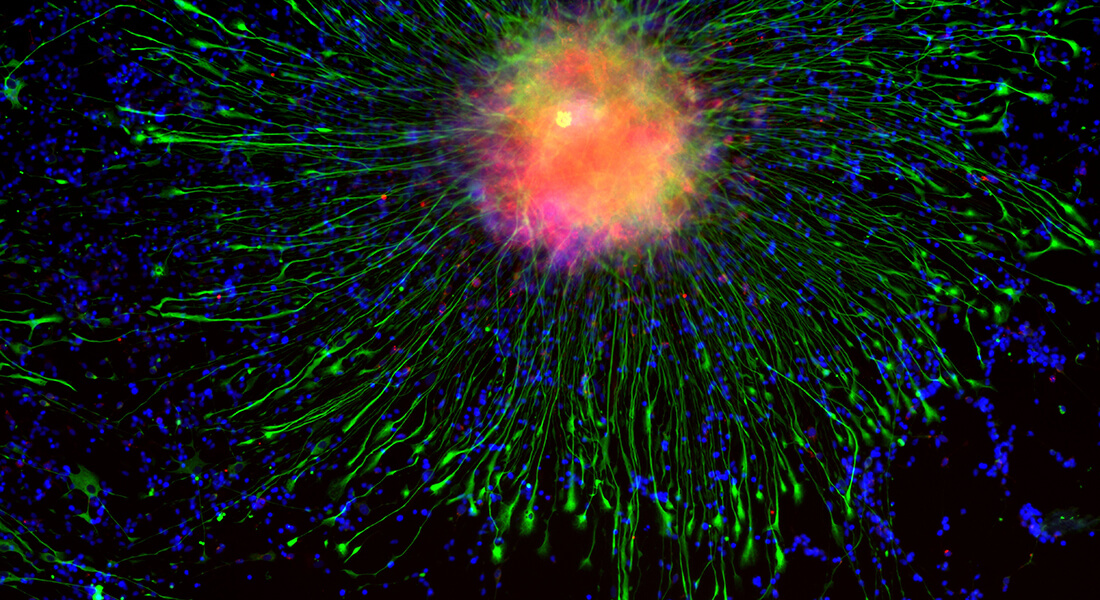
The glymphatic system is responsible for clearing toxic metabolites from the brain during sleep. This network of perivascular spaces lined by astrocytic end feet enables the bulk fluid movement of cerebrospinal fluid into, and interstitial fluid through the brain. Decreased glymphatic function, and thus decreased clearance of waste, has been associated with age, Alzheimer’s disease, diabetes, hypertension, traumatic brain injury, chronic high ethanol intake, chronic unpredictable stress, bacterial meningitis, and, preliminarily, pain.
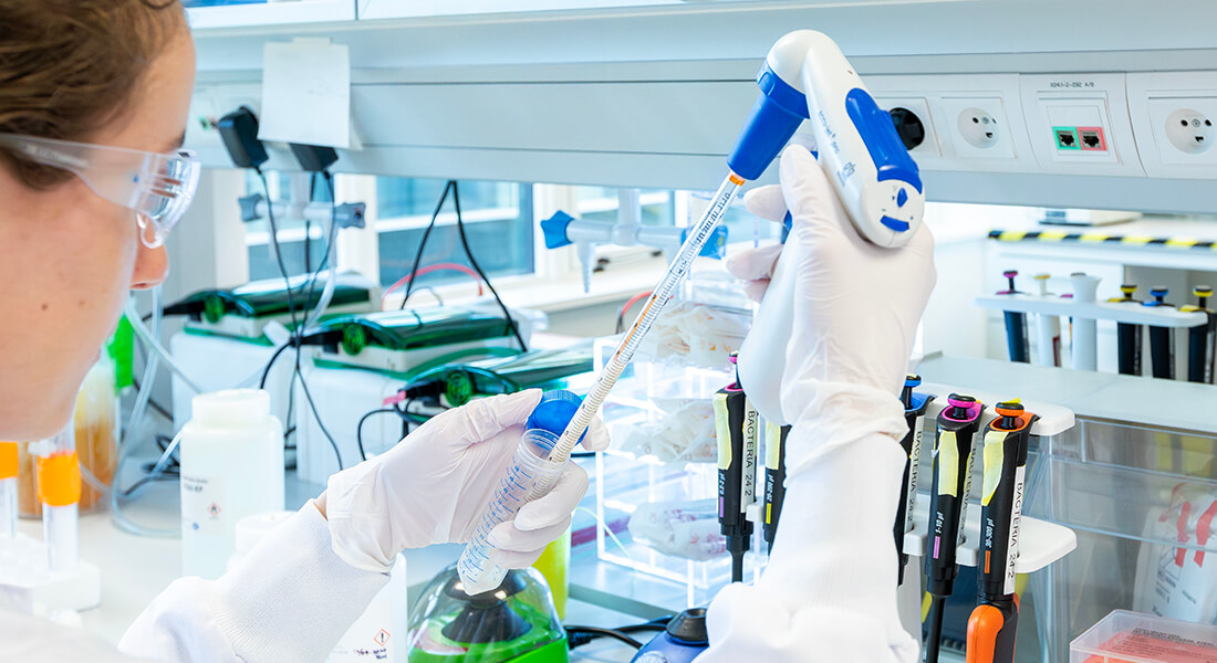
The majority of these conditions that exhibit glymphatic dysfunction are also associated with increased neuroinflammation characterized in part by increased inflammatory cytokines and reactive gliosis. The main goal of this master’s proposal is to understand how neuroinflammation may alter the structure of the glymphatic system, either in the perivascular or interstitial space, leading to a reduction in flow. This project will involve a combination of live-animal surgery, ex vivo and in vivo imaging, in vivo microdialysis, immunohistochemistry and more.<span data-ccp-props="{" 201341983":0,"335559739":160,"335559740":259}"="201341983" :0,"335559739":160,"335559740":259}""=":0," 335559739":160,"335559740":259}"""="335559739" :160,"335559740":259}""""=":160," 335559740":259}"""""="335559740" :259}""""""=":259}" """"""="" """"""="">
This project has a special emphasis on chronic pain, a debilitating condition with no effective, non-addictive treatment. We use two pre-clinical models of chronic neuropathic pain: sparse nerve injury and spinal chord injury. Our goal is to understand glymphatic dysfunction in chronic pain, and potentially develop therapeutics to increase glymphatic flow after development of neuroinflammatory pathology.<span data-ccp-props="{" 201341983":0,"335559739":160,"335559740":259}"="201341983" :0,"335559739":160,"335559740":259}""=":0," 335559739":160,"335559740":259}"""="335559739" :160,"335559740":259}""""=":160," 335559740":259}"""""="335559740" :259}""""""=":259}" """"""="" """"""="">
The project will take place in the Center for Translational Neuromedicine (CTN) at the University of Copenhagen and the University of Rochester Medical Center. CTN is an international research group of more than 50 people, with collaborations that span the globe, providing exposure to a wide array of scientific questions centered around glial cells. CTN features state-of-the-art facilities and equipment, joint weekly journal clubs and focus groups between Copenhagen and Rochester, and integrative training among undergraduates, graduate students, postdocs and faculty. By integrating international teams and training levels, trainees are exposed to all levels of science from benchwork to science writing to project development. <span data-ccp-props="{" 201341983":0,"335559739":120,"335559740":240}"="201341983" :0,"335559739":120,"335559740":240}""=":0," 335559739":120,"335559740":240}"""="335559739" :120,"335559740":240}""""=":120," 335559740":240}"""""="335559740" :240}""""""=":240}" """"""="" """"""="">
This project is a collaboration between Dr. Lauren Hablitz, an expert in glymphatic function, and Dr. Maiken Nedergaard, an expert in glial biology, pain, and discoverer of the glymphatic system. As such, this project will involve research done in Rochester, New York, USA. The lab language is English, and international as well as Danish students are welcome. <span data-ccp-props="{" 201341983":0,"335559739":120,"335559740":240}"="201341983" :0,"335559739":120,"335559740":240}""=":0," 335559739":120,"335559740":240}"""="335559739" :120,"335559740":240}""""=":120," 335559740":240}"""""="335559740" :240}""""""=":240}" """"""="" """"""="">
Please send your application including a short CV and a short letter of motivation to Dr. Hablitz or Dr. Nedergaard.<span data-ccp-props="{" 201341983":0,"335559739":120,"335559740":240}"="201341983" :0,"335559739":120,"335559740":240}""=":0," 335559739":120,"335559740":240}"""="335559739" :120,"335559740":240}""""=":120," 335559740":240}"""""="335559740" :240}""""""=":240}" """"""="" """"""="">
Department of Neurology<span data-ccp-props="{" 201341983":0,"335559739":160,"335559740":240}"="201341983" :0,"335559739":160,"335559740":240}""=":0," 335559739":160,"335559740":240}"""="335559739" :160,"335559740":240}""""=":160," 335559740":240}"""""="335559740" :240}""""""=":240}" """"""="" """"""="">
Center for Translational Neuromedicine <span data-ccp-props="{" 201341983":0,"335559739":160,"335559740":240}"="201341983" :0,"335559739":160,"335559740":240}""=":0," 335559739":160,"335559740":240}"""="335559739" :160,"335559740":240}""""=":160," 335559740":240}"""""="335559740" :240}""""""=":240}" """"""="" """"""="">
University of Rochester Medical Center<span data-ccp-props="{" 201341983":0,"335559739":160,"335559740":240}"="201341983" :0,"335559739":160,"335559740":240}""=":0," 335559739":160,"335559740":240}"""="335559739" :160,"335559740":240}""""=":160," 335559740":240}"""""="335559740" :240}""""""=":240}" """"""="" """"""="">
Streptococcus pneumoniae meningeal infection promotes gliosis in the brain parenchyma. (A–D) Confocal microscopy of coronal brain sections obtained from S pneumoniae–infected (A, C, upper panel) and control rat brains immunolabeled for glial fibrillary acidic protein (GFAP; A, C; red), microglial/macrophage ionized calcium‐binding adapter molecule 1 (Iba1; B, D; yellow), and neutrophil myeloperoxidase (MPO; A; green) and counterstained with 4',6‐diamidino‐2‐phenylindole (DAPI; blue). (A,C) In infected animals, astrogliosis was observed in brain areas with bacterial proliferation and recruitment of neutrophils such as the meninges and the lateral ventricle (LV), and also in areas where bacteria and neutrophils were not detected, namely the hippocampus (Hpc; C, arrow). (A) Direct cell–cell contacts between astrocytes and neutrophils were observed in the meninges and in the lateral ventricle. (C, lower panel) Astrocytes were not activated in the meninges, lateral ventricle, and hippocampus of control animals. Activated and hypertrophic microglia were observed in the meninges of infected rats (B, D, yellow inset, arrows), in direct contact with infiltrating neutrophils. There was also a massive presence of meningeal macrophages (B, D, blue inset, arrows), directly interacting with neutrophils (B, arrows). In sharp contrast, there was no sign of microglial activation in control animals (D, lower panel, arrows), which had only sporadic macrophages present in the meninges. (E–G) Quantification of GFAP immunoreactivity in the cortex (CTX; E) and hippocampus (F) and Iba1+ immunoreactivity in the cortex (G; unpaired t‐test with Holm–Šídák multiple comparison adjustment, *p < 0.05,
**p < 0.01, ***p < 0.001). Error bars represent ± standard deviation, n = 8 in each group. Scale bars: 10μm. Lm = leptomeninges; SP001 = S pneumoniae strain 001. Annals of Neurology, Volume: 90, Issue: 4, Pages: 653-669, First published: 16 August 2021, DOI: (10.1002/ana.26186).
Together, both facilities house several micro and macroscopy units, SPECT/PET/μCT scanner and 9.4T ultra-high field magnetic resonance imaging (MRI) equipment along with a large variety of coils and peripheral devices. By using the most optimal setup for your experiments, you will have a chance to generate new and valuable data, analyze it, and draw important neuroscientific conclusions.
Project: The project will employ advanced imaging techniques, such as MRI and μCT, to visualize and quantify glymphatic function in animal models under different anesthesia protocols. By comparing the glymphatic activity levels and patterns between awake and anesthetized states, the study seeks to elucidate how anesthesia affects the efficiency and dynamics of waste clearance in the brain.
By joining us, you would help us to answer the question of how far the results found under anesthetic protocols impair the translational value for glymphatic MRI. Findings from this research endeavor would inform clinical practices involving anesthesia administration in neuroscience research and medical procedures, ultimately advancing our understanding of brain health and disease mechanisms. The exact project outline would be tailored to the specific skills and background of each candidate.
Candidate: The candidate should possess skills in small animal handling, and be of medical, biomedical, biophysical, or biological background. Skills in wet-lab or basic coding in Matlab or Python are superlative but not substantial. The candidate would receive training in MRI and μCT, and would be required to communicate and coordinate the experiment in a team-oriented manner.
Contact: Ryszard Gomolka, Assistant Professor at the Center for Translational Neuromedicine and MRI Core Facility. (ryszard.gomolka@sund.ku.dk)
Please send your1 page CValong with a short1/2 pagemotivation letter describing your to date background and interest.
Migraine affects up to 1 out of 10 and is characterized by a unilateral throbbing headache which can be followed by nausea, vomiting, photo and phonophobia, and exacerbation of headache during physical activity. In about one third of migraine patients, the migraine headache is preceded by transient neurological symptoms lasting 5-60 minutes.
Headache disease models are notoriously tricky to characterize in animal models, since specific biomarkers are lacking, and behavioral assays are only somewhat able to detect the subtle changes migraine attacks might inflict. Just think about how in humans, migraine diagnostics is 100% based on a patient interview, and therefore in clinical settings based on the patient’s subjective experience. This is of course impossible to do in a rodent model.
However, the technological and computational advancement in science has started to change this. We are planning to use tools such as optogenetics, fiber photometry and behavioral sequencing (e.g., Deep Lab Cut) to generate a behavioral assay for migraine with aura in mice where the effect of putative migraine abortive and preventive drugs can be tested.
We are seeking a master student with an interest in translational neuroscience to develop an animal preparation where we use transgene mice to induce migraine with aura using optogenetics, and where we quantify migraine attacks with genetically encoded calcium indicators. All this is to be done in a freely moving awake animal.
About the candidate:
Preferably you have experience with small animal handling, and if you already have FELASA certification that is a plus. The candidate will be trained in transgenic models, optogenetics, fiber photometry and behavioral sequencing. Any prior experience in any of these fields is an advantage for the candidate.
Contact:
Please write an email with your letter of interest and including a 1-page CV to post doc Martin Kaag Rasmussen, Center for Translational Neuromedicine at martin.rasmussen@sund.ku.dk.
Are you interested in playing a major role in deciphering:
How does this fatal neurodegenerative disease, ALS, progress with aging?
What happens to the upper and lower motor pathways during the disease progression?
How the glymphatic system is modulated during the disease progression?
If you are curious to learn and answer these important questions in your thesis project this is the project for you!
Why is it important to study Amyotrophic lateral sclerosis (ALS)?
Amyotrophic lateral sclerosis (ALS) is an incurable condition, characterized by degeneration of upper motor neurons (in the brain) and lower motor neurons (in the brainstem and spinal cord) leading to weaker muscles, paralysis and eventually death within 2-5 years. According to Lancet Neurology study, about 3.4 billion individuals were living with neurological conditions worldwide equating to 43.1% global population (Steinmetz et al, 2021) of which, over 331,000 individuals were suffering from ALS (Vos et al, 2017). Even though major symptoms of ALS constitute motor dysfunction, up to 50% patients develop cognitive impairment, 13% develop frontotemporal dementia (FTD) and the most common mutation occurs in superoxide dismutase (SOD1) gene. Riluzole is the only effective and clinically used drugs globally to reduce glutamate mediated excitotoxicity and thus prolongs lifespan to approximately 3 months. However, the drug comes with side-effects which further compromises the quality of life of ALS individuals.
What are we lacking in order to find an appropriate treatment for ALS?To find an effective treatment for ALS, we first need to understand what is happening to the neurons and glial cells at network level before the onset of disease and during the disease progression in the brain as well as the spinal cord. Revealing the progression of the disease at neuroglial network level with help understanding its complex mechanism.
Here you will use an SOD1 mouse model of ALS. These mice carry mutations in the SOD1 gene which causes motor neuron degeneration and mimic various aspects of ALS in humans. You will use in vivo imaging (such as fiber photometry and 2-photon imaging) and in vitro assays to reveal the mechanistic changes at neuroglial network level in ALS.
Candidate: You are a motivated student with an interest in neurodegenerative diseases and interest in how the brain as well as the spinal cord work. You are curious to learn new skills and a team player. You are required to have a Felasa certificate and can communicate in English.
Contact: Send your CV to Jaspreet Kaur Daniil, Assistant Professor at Center for Translational Neuromedicine, email: jaspreet.kaur@sund.ku.dk
Centre for Translational Neuromedicine, University of Copenhagen
About the project
Exploring how the brain dynamically regulates blood-brain barrier (BBB) permeability during learning will have profound implications for understanding memory formation and brain plasticity. This MSc project will investigate how learning alters BBB brain-wide permeability using a novel transgenic mouse model, molecular tools and advanced imaging techniques.
Students will perform behavioural experiments such as contextual fear conditioning. Brains will then be processed using tissue clearing techniques (e.g. PEGASOS, iDISCO) and whole-brain imaging using light-sheet microscopy to visualize and quantify the spatial distribution of bloodborne biomolecules that cross the BBB in response to memory formation.
Techniques Involved
In vitro cell biology: Production of viral tools for studying vasculature function (optional)
Behavioral neuroscience: Fear learning paradigm (contextual fear conditioning)
Image analysis: 3D reconstruction and quantification of BBB permeability
Details
Supervisors: Prof. Hajime Hirase & Dr. Antonis Asiminas
Location: Centre for Translational Neuromedicine, University of Copenhagen
Start Date: Flexible
Duration: 9 months
About you
We are looking for highly motivated MSc students with an interest in neuroscience, imaging, and blood-brain barrier dynamics. Ideal candidates should have:
Experience or strong interest in neurobiology and imaging techniques
Experience in in vitro cell biology techniques (optional)
FELASA certification for working with laboratory animals (strongly preferred)
Good communications skills in English
MSc students with in vitro cell biology experience who do not holdFELASA are also encouraged to reach out to discuss variations of this project not involving animal experiments.
About us
We are based in Panum, at Centre for Translational Neuromedicine. We are an international research team focusing on the interactions between neurons, glia and vasculature in health and disease.